This section of our website will be dedicated to show you the many different views of how RSD/CRPS can affect the body. In this RSD photo gallery, you will view photos that show many of the different types of symptoms that are associated with RSD/CRPS such as edema, discoloration of the skin, skin ulcers/lesions, infections, deformities of the extremities and digits. You will also see some before and after treatment photos along with some Thermography photos.
We know that these photos are disturbing to view. This does not mean that every RSD/CRPS patient will end up like this. We are just showing what some patients have been through with their journey of this complex disease we call RSD/CRPS.
Take a few moments to browse through the gallery below. If you have any questions, please contact us at: utopia331965@gmail.com .
RSD of The Left Foot Pictures
|

|

|
")
|

|
|
Leg and Foot Deformity-Discoloration Picture #1 Onset of RSD 12-7-1985.
|
Left Foot Deformity Picture #2
Three years after onset and before surgery. The toes became fused over a three year span. 12-7-1988.
|
Left Foot Deformity Picture #3
Post-Op Fusion of Great Toe
12-14-1988.
|
Left Foot Deformity Picture #4 Five years after onset of RSD 1990.
|
|

|

|

|
|
Left Foot Deformity Picture #5
Eight years after onset of RSD 1993.
|
Left Foot Deformity Picture #6
Seventeen years after onset of RSD. Please note toenail on left great toe. 7-11-2002.
|
Left Foot Deformity Picture #7
Seventeen years after onset of RSD. Please note after removal of toenail on left great toe. 7-12-2002.
|
RSD and Infection Pictures
|

|

|

|
|
RSD Infection Picture #1
RSD of the left foot. The infections started in the 22nd year of having RSD.
|
RSD Infection Picture #2
The infections lasted for over a year and half. Ultimately the left foot and leg above the knee were amputated in August of 2008. This was 23 years after onset of RSD due to a car accident.
|
RSD Infection Picture #3 The patient lost the use of the limb after his first surgery to fuse the great toe in 1988. Three years after the onset of RSD.
|
Skin Lesion and Skin Ulcer Pictures
|
")
|

|

|
|
Skin ulcers before and after treatment of epidural, regional nerve blocks, and I.V. Mannitol, showed marked improvement of skin ulcers.
|
Skin ulcers caused from application of ice.
Five years of daily self-applied cryotherapy (application of ice) resulted in the breakdown of skin, cold blisters, and damage to sensory nerves.
|
Skin ulcers after treatment.
Eleven days after discontinuation of cryotherapy, treatment of epidural, regional nerve blocks, and I.V. Mannitol, showed marked improvement of the bulbous lesions.
|
|

|

|

|
|
Skin lesions from Venipuncture CRPS II Injury.
Venipuncture CRPS II, five months after a blood test, resulted in neuroinflammatory bulbous lesions.
|
Ulcers from Venipuncture CRPS II Injury.
Lesions became ulcerated.
|
Venipuncture CRPS II lesions after treatment.
The lesion healed after treatment using I.V. Mannitol and I.V. immunoglobulin.
|
RSD of The Hand Pictures
|

|

|
|
Hand Deformity Picture #1
RSD of the right hand with deformity.
|
Hand Deformity Picture #2
The wrist and digits became fused. The patient was not able to move the wrist or digits.
|
|

|

|
|
Hand Deformity Picture #3
RSD of the right hand after multiple Carpal Tunnel surgeries.
|
Hand Deformity Picture #4
Deformity was caused by multiple carpal tunnel surgeries. This patient also had the (little finger) 5th digit amputated.
|
Thermography Pictures
|

|

|

|
|
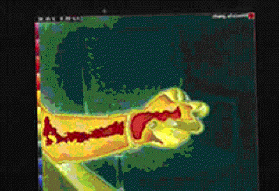
Venipuncture CRPS II Thermography
Venipuncture CRPS II Damage to the Median Nerve.
|
Thermography of the Right Hand (a.k.a.) "Boxers Hand Deformity."
CRPS of seven years duration due to right hand injury. Two years of unsuccessful operations at right carpal tunnel, and five years of immobilization of hand have resulted in "Boxers Hand Deformity" and ultimate amputation.
|
Virtual Sympathectomy Thermography.
"Virtual sympathectomy" secondary to repeated stellate ganglion nerve blocks leading to permanent sympathetic nerve damage and hyperthermia (heat leakage) in upper extremities. The ITI spared the patient from further sympathetic nerve blocks.
|
|

|

|

|
|
Thermography of an Electircal Injury Patient.
Central hyperthermic areas of entrance and exit in electrical injury. The permanent hyperthermic damage is surrounded by vasoconstrictive hypothermia. Only after increasing the thermal sensitivity (right) the lesions were identified. This "button hole" sign is exclusively seen in electrical injury.
|
Thermography of the face and hands after Sympathectomy.
|
Thermography of the Hands (After Virtual Sympathectomy).
More than two dozen stellate ganglion blocks to each side have damaged enough sympathetic nerve to cause permanent hyperthermia as the manifestation of virtual sympathectomy. Further blocks have no diagnostic or therapeutic value.
|
|

|

|

|
|
RSD Neuroma Exploration Thermography.
CRPS nerve damage to right toes after "neuroma exploration". The sympathectomy did nothing for the pain. ITI spared the patient from the scheduled chemical sympathectomy. The left foot showed compensatory hypothermia after sympathectomy.
|
Cryotherapy Thermography
Bilateral Thermogram shows marked hypothermia due to vasoconstriction in the cryotherapy area.
|
AVM-RSD Thermography
A previously undiagnosed right leg arteriovenous malformation (AVM)over 27mm deep, complicated by CRPS (RSD) . ITI identified the deep lesion and spared the patient from the scheduled sympathectomy. Vascular surgery corrected the condition.
|
Anatomy Sketches
In this section of our website you will find some anatomy sketches of the Sympathetic Nervous System, Parasympathetic Nervous System, Peripheral Nervous System, The Circulatory System and many other sketches.
We hope that these anatomy sketches will be helpful to you.
Please click on the link below to view these anatomy sketches in PDF-format.
ANATOMY SKETCHES